Endodontic surgery
Apicoectomy (Periapical Surgery)
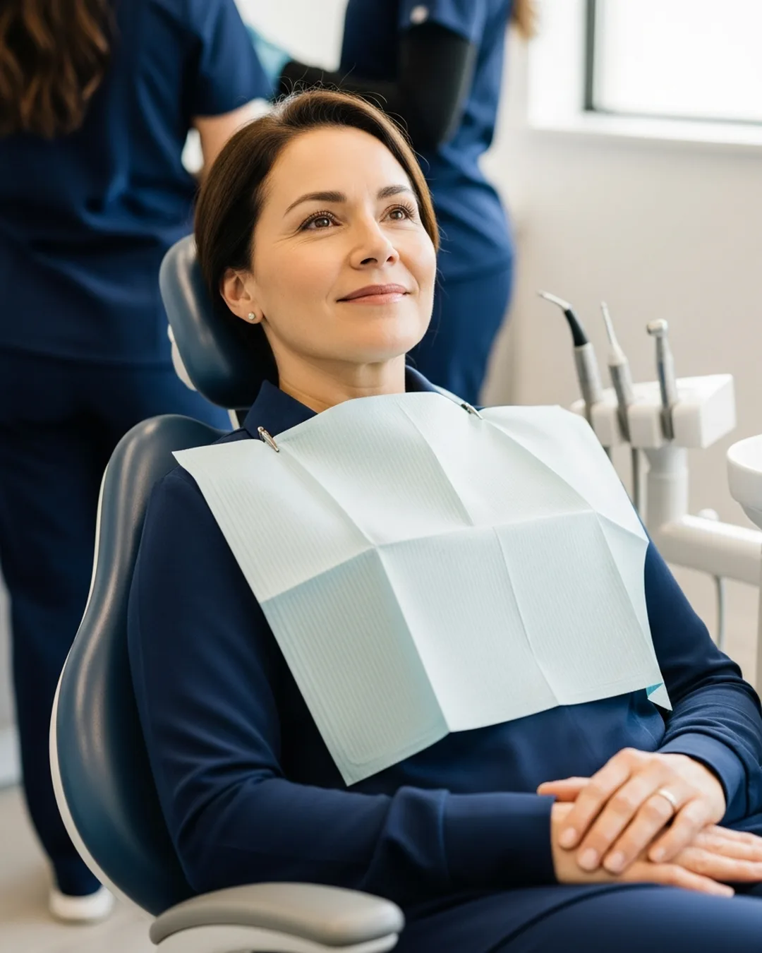
An apicoectomy is the option that keeps a treated tooth in place when infection at the root has not cleared. Your endodontist plans it from a 3D CBCT scan and works under a surgical microscope. You feel pressure but no pain under local anesthesia, and you are home resting within a couple of hours.
Find Your Nearest Office →An apicoectomy (a small surgery on the tip of a tooth's root) clears infection that lingers after a root canal. The endodontist removes the inflamed tissue and the root tip, then seals the end. In a 2025 review of 22 studies, 89% of treated teeth were successful (Ko and colleagues, Restorative Dentistry and Endodontics, 2025). Most people are back to normal activities within a few days. It's usually the final step to save the tooth.
It’s the microsurgical option for saving a tooth when infection sits at the very tip of the root, beyond the reach of treatment from inside the tooth. You may also hear it called periapical surgery or endodontic microsurgery. At Mid-Florida Endodontics, an endodontist performs it through a tiny opening in the gum, working under a surgical microscope so only the affected root tip is treated. It’s typically considered after a root canal or retreatment hasn’t fully cleared the problem, and it lets you keep your natural tooth.
When Is an Apicoectomy Needed?
An apicoectomy is not the first line of treatment, it is indicated when root canal treatment has been performed but a persistent problem cannot be resolved through the crown of the tooth alone. Common indications include:
Persistent Periapical Infection After Root Canal or Retreatment
A periapical lesion (an area of infection and bone damage at the tip of a tooth’s root, visible on X-rays) that fails to heal, or continues to grow, after completed root canal treatment or retreatment indicates that bacteria or infected tissue persists at the root tip that cannot be reached through conventional canal instrumentation.
Canal Anatomy That Cannot Be Retreated From the Crown
Severely calcified canals, a post and core that cannot be safely removed, or an instrument separated at the root tip may make non-surgical retreatment impossible or carry too high a risk of tooth damage. Surgical access from the root end bypasses these obstacles entirely.
Biopsy of a Suspicious Periapical Lesion
Periapical lesions are almost always inflammatory in nature, but occasionally, a lesion that does not respond as expected to treatment requires biopsy to rule out non-inflammatory pathology. Apicoectomy provides both surgical treatment and the tissue sample needed for pathological analysis.
Anatomical Factors Near the Root Tip
Accessory canals, isthmuses, and lateral canals in the apical third of the root may harbor bacteria that are unreachable by instrumentation from above. Resecting the apical few millimeters of the root removes this reservoir of infection entirely.
Why the Surgical Microscope Changes Apicoectomy Outcomes
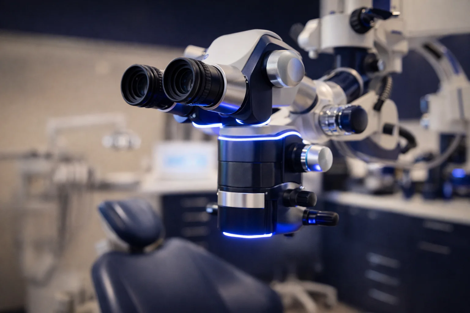
Traditional apicoectomy, performed without magnification, required a larger surgical access, less precise root-end preparation, and produced more variable outcomes. Under the surgical microscope at high magnification, our specialists operate in a field of millimeter-level precision: resecting the exact amount of root, inspecting the cut root surface for isthmi and lateral canals. Preparing and sealing the root end with accuracy impossible to the naked eye. Research comparing microscope-assisted endodontic microsurgery to traditional surgery shows significantly higher success rates for the microscopic technique.
What Happens During an Apicoectomy?
The procedure is performed under local anesthesia in our office. Most patients are surprised by how manageable it is. Here is exactly what happens:
Pre-Surgical CBCT (cone-beam CT, a low-dose 3D X-ray that lets us see the tooth and bone from every angle) Imaging
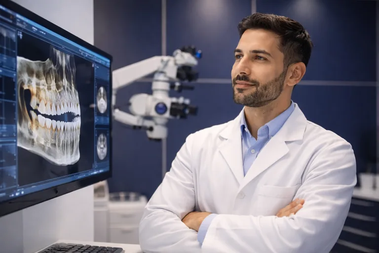
A 3D CBCT scan maps the root tip position relative to critical anatomy, the inferior alveolar nerve, maxillary sinus, and adjacent root apices. This information determines the precise angle and depth of surgical access and prevents injury to surrounding structures.
Local Anesthesia
The area is thoroughly numbed. You will feel pressure during the procedure but no pain. We take all the time needed to ensure you are fully comfortable before proceeding.
Gum Tissue Incision and Flap Reflection
A small incision is made in the gum tissue near the affected root. The tissue is gently reflected to expose the bone and root tip below. The incision is designed to heal with minimal scarring and is typically small enough that sutures are removed within a week.
Removal of Infected Tissue and Root Tip Resection
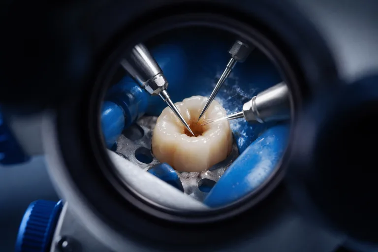
The infected tissue surrounding the root tip is carefully removed. The apical 2-3 millimeters of the root are then resected, removing the portion of the root that harbors the greatest concentration of accessory canals and potentially infected debris. The resected root end is inspected under the microscope for any isthmi, cracks, or untreated anatomy.
Root-End Preparation and retrofill (sealing the cut root end from the outside during apicoectomy surgery)
A small cavity is prepared in the cut root surface using an ultrasonic tip under microscope visualization. This cavity is then filled with a biocompatible material, typically mineral trioxide aggregate (MTA), a biocompatible dental cement used to seal the root end during apicoectomy surgery . This Seals the root end and creates a bacteria-tight barrier. The precise preparation and sealing under magnification is the feature that distinguishes modern microsurgery from older techniques.
Closure and Healing
The tissue flap is repositioned and sutured closed. A bioresorbable membrane may be placed to support bone regeneration if significant bone loss was present. Sutures are typically removed at a follow-up appointment 5-7 days later.
What Is Recovery Like After an Apicoectomy?
Peak Swelling
Swelling and bruising in the area are expected, this is a normal surgical response, not a sign of complication. Take medications as prescribed or recommended by your provider. Eat soft foods and avoid the surgical area.
Suture Removal
Swelling begins to subside. A follow-up appointment removes sutures. Most patients are comfortable returning to normal activities within a few days, though strenuous exercise should be avoided for about a week.
Bone Healing Confirmed
Periapical bone healing is monitored with follow-up imaging. Full bone regeneration takes several months to a year. Successful healing is confirmed when the lesion has resolved on imaging.
Important: Ice packs applied to the outside of the face can help manage swelling in the first 24-48 hours after surgery, heat and ice may both be used as directed by your provider. Always follow your provider’s specific post-operative instructions. Contact us if you develop significant worsening of swelling after day 3, fever, or signs of wound dehiscence.

What Are the Success Rates for Apicoectomy?
Microsurgical Apicoectomy: High Success in Appropriate Cases
A meta-analysis published in the Journal of Endodontics found that endodontic microsurgery, apicoectomy performed under microscope magnification with ultrasonic root-end preparation and MTA (mineral trioxide aggregate, a biocompatible cement used to seal the root tip during surgery) retrofill, achieves success rates of approximately 92-94% at follow-up, significantly higher than outcomes reported for older macro-surgical techniques. Appropriate case selection, 3D pre-surgical imaging, and microscope-guided precision are the primary drivers of these outcomes. (Setzer FC, et al. Outcome of endodontic surgery: a meta-analysis of the literature. J Endod. 2010;36(11):1757-1765.)
Frequently asked questions
What is an apicoectomy?
Is an apicoectomy painful?
How long does an apicoectomy take?
How long is the recovery after an apicoectomy?
Why would I need an apicoectomy instead of another root canal?
How successful is an apicoectomy?
Will I need a new crown after an apicoectomy?
How soon after an apicoectomy can I eat normally?
Persistent infection after a root canal?
When a root canal was not enough, periapical surgery removes the infection at the source and saves the tooth.
Schedule a surgical consultationSpecialist Apicoectomy (Periapical Surgery) near you
-
Apicoectomy (Periapical Surgery) in Clermont
-
Apicoectomy (Periapical Surgery) in Daytona Beach
-
Apicoectomy (Periapical Surgery) in Dr. Phillips, Orlando
-
Apicoectomy (Periapical Surgery) in Kissimmee
-
Apicoectomy (Periapical Surgery) in Lake Nona, Orlando
-
Apicoectomy (Periapical Surgery) in Longwood
-
Apicoectomy (Periapical Surgery) in Maitland
-
Apicoectomy (Periapical Surgery) in Mount Dora
-
Apicoectomy (Periapical Surgery) in Orange City
-
Apicoectomy (Periapical Surgery) in Waterford Lakes, Orlando
-
Apicoectomy (Periapical Surgery) in Winter Garden