The root canal’s fearsome reputation was earned decades ago, when hand files were rigid, X-rays were flat and imprecise, anesthesia was less predictable, and the procedure could take multiple lengthy visits. That era of endodontics is over.
Today, a root canal performed by an endodontic specialist is a precisely engineered procedure supported by imaging technology, magnification, and instrumentation that simply did not exist a generation ago. Understanding what these tools do, and why they matter, helps explain why patients at Mid-Florida Endodontics so consistently describe their experience as far easier than they expected. The goal of every one of these advances is the same: to help the endodontist save the natural tooth comfortably and predictably.
Then vs. Now: How Technology Transformed Root Canals
Root Canals: Past
- Flat 2D X-rays, limited anatomy view
- Naked eye or basic loupes only
- Rigid stainless steel hand files
- Multiple appointments common
- Canal length estimated by X-ray
- Anesthesia less predictable
- Higher rate of missed canals
Root Canals: Today
- 3D CBCT imaging, full anatomy in three dimensions
- Surgical microscope, high-power magnification
- Flexible NiTi rotary instruments
- Most cases completed in one visit
- Electronic apex locators, precise to fractions of mm
- Computer-assisted anesthesia delivery
- Ultrasonic instrumentation for multiple purposes
The Technologies That Define Modern Endodontics
3D Cone-Beam CT (CBCT) Imaging
Before modern endodontics, dentists worked from flat 2D X-rays that showed teeth from a single angle, causing structures to overlap and hiding critical anatomy. CBCT imaging creates a full three-dimensional model of the tooth and surrounding structures, revealing the number, length, curvature, and position of every root canal before a single instrument is placed.1 Studies show this added detail changes how endodontists plan treatment in a meaningful share of complex cases.1 At Mid-Florida Endodontics, CBCT imaging is used routinely on the vast majority of patients because the diagnostic information it provides is too valuable to forgo.
Patient benefit: Fewer surprises, more accurate treatment, better outcomes
Surgical Operating Microscope
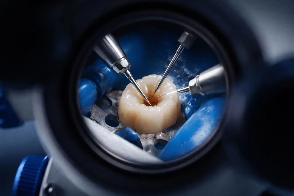
The surgical microscope provides high-power magnification with coaxial illumination (lighting that comes from exactly the same angle as the line of sight through the microscope, eliminating shadows in the surgical field), so light enters from precisely the direction the endodontist is looking.8 Under this magnification, the endodontist can identify extra canals invisible to the naked eye, detect hairline cracks in tooth structure, and confirm complete removal of pulp tissue. One clinical series found that adding the operating microscope and microendodontic instruments raised the rate at which a commonly missed second canal in upper first molars was located to about 93 percent.2 The same magnification lets the specialist perform precision microsurgery, and a meta-analysis of surgical outcomes reported markedly higher success when microsurgical techniques were used compared with traditional root-end surgery.3 At Mid-Florida Endodontics, the microscope is used on every case.
Patient benefit: More canals found and treated, fewer treatment failures
Computer-Assisted Anesthesia (The Wand)
Traditional dental injections deliver anesthetic in a rapid bolus, the discomfort patients associate with “the needle.” The Wand system uses computer-controlled delivery to release anesthetic slowly and steadily at a rate that the tissue can accommodate more comfortably. A randomized clinical trial comparing a computer-controlled delivery system with a conventional injection found lower reported injection pain with the computer-controlled approach.4 Most patients describe the experience as far more comfortable than they expected, and this technology is one of the most direct contributors to the improvement in patient comfort during modern root canal treatment.
Patient benefit: Significantly more comfortable numbing experience
Nickel-Titanium (NiTi) Rotary Instruments
Earlier root canal instruments were made from stainless steel, relatively rigid files that had to be hand-worked in and out of curved canals. Nickel-titanium alloy files are highly flexible, conforming to natural canal curves with less risk of straightening or transportation than steel instruments. Driven by a precision motor at controlled torque and speed, NiTi rotary instruments help the endodontist clean and shape canals efficiently and safely, and a review of canal-preparation evidence concluded that shaping outcomes with these instruments are mostly predictable in trained hands.5 They also shorten the cleaning and shaping step compared with the older hand-filing technique.
Patient benefit: Faster treatment, better canal shaping, reduced procedural risk
Electronic apex locator
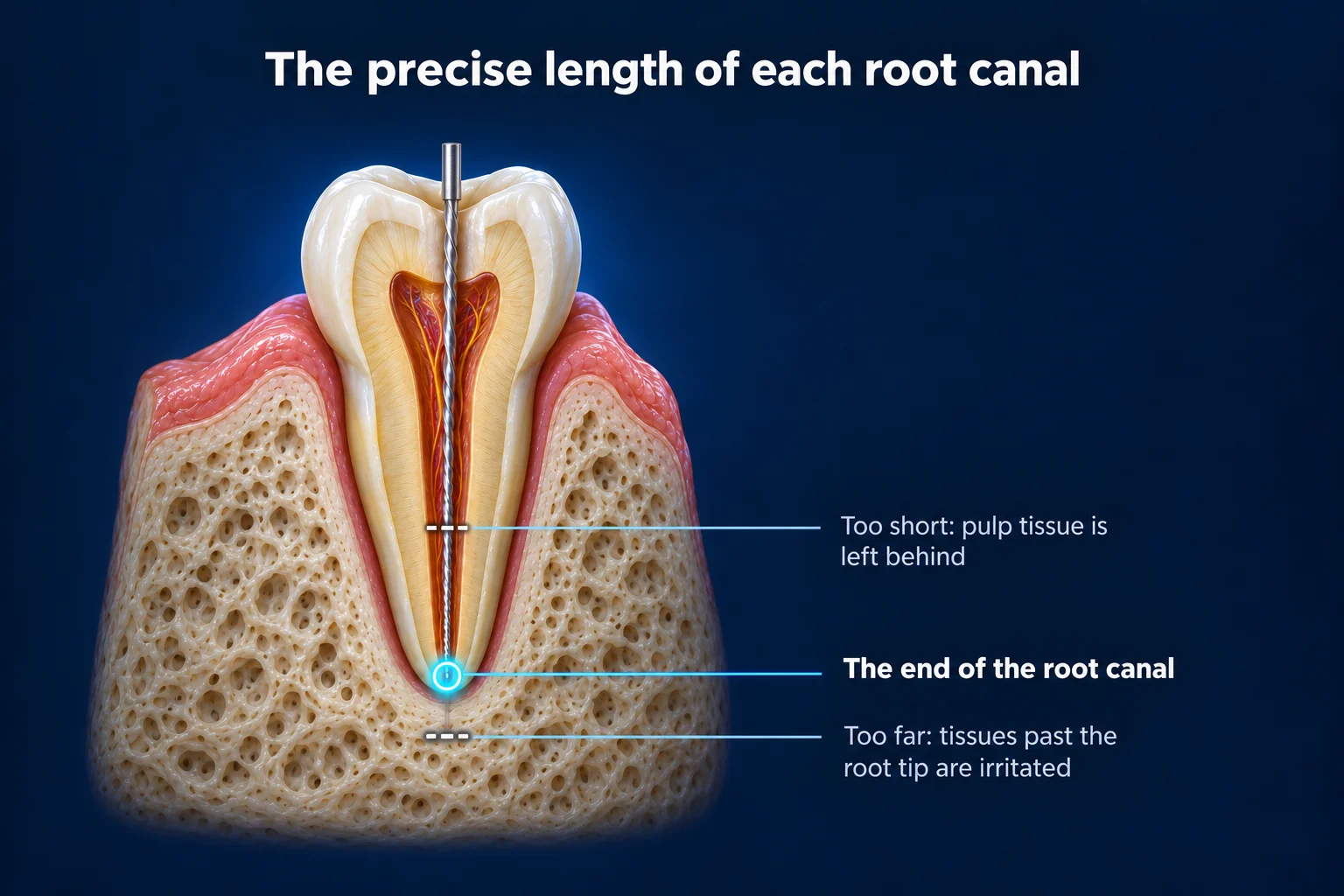
Knowing the precise length of each root canal is critical: an instrument too short, and pulp tissue is left behind; an instrument too far, and the periapical (relating to the area surrounding the very tip of a tooth’s root) tissues are irritated. Historically, this relied on X-ray estimation. Electronic apex locators use electrical impedance to detect the position of the instrument tip relative to the end of the root canal, in real time and without additional radiation exposure. A systematic review and meta-analysis of multiple device generations found that electronic apex locators reliably determine working length, which is why endodontists depend on them.6
Patient benefit: Precise treatment length, no under- or over-preparation
Ultrasonic Instrumentation
Ultrasonic tips vibrating at high frequency serve multiple functions in modern endodontics: activating irrigating solutions to enhance disinfection throughout the canal system, troughing (using an ultrasonic instrument to carefully remove a thin groove of tooth structure to locate hidden root canal openings) the pulp (the soft living tissue inside the tooth, containing nerves and blood vessels) chamber (the hollow space inside the crown of the tooth that houses the nerve and blood vessels) floor to locate hidden canal orifices, and removing posts and other restorative materials during retreatment, including the retrieval of separated instruments. A systematic review and meta-analysis found that activating the irrigating solution, including with ultrasonic energy, improves how thoroughly debris and the smear layer (a thin film of residue left on the canal wall after cleaning) are cleared across most of the canal, which is why activation is recommended throughout the cleaning step.7 Under microscope guidance, ultrasonic instrumentation helps the endodontist make many previously difficult procedures routinely achievable.
Patient benefit: More thorough disinfection, better retreatment capability
Advanced Obturation Systems
Sealing the cleaned canals completely, without voids that could harbor residual bacteria, is the final critical step of root canal treatment. Modern obturation techniques and materials allow endodontists to achieve a dense, three-dimensional seal of the entire canal system using gutta-percha (a natural rubber-like material used to fill and seal root canals after treatment, the standard material for well over a century) and advanced biocompatible sealers. The goal is a leak-resistant seal that helps prevent bacterial recontamination over the long term when it is combined with a properly placed permanent crown.
Patient benefit: Better long-term seal, reduced risk of reinfection
These technologies have been studied individually rather than as a single bundle. A review of root canal preparation in the Journal of Endodontics concluded that, with appropriate training, shaping outcomes using nickel-titanium rotary instruments are mostly predictable, while noting that randomized clinical trials are still needed to compare long-term outcomes against earlier hand techniques.5 Imaging and magnification carry their own evidence: a systematic review found that cone-beam CT changes endodontic treatment planning in a meaningful share of complex cases,1 and a meta-analysis of surgical outcomes reported substantially higher success with microsurgical (microscope-based) techniques than with traditional root-end surgery.3 Taken together, the evidence supports that each modern tool, in the hands of an endodontic specialist, contributes to more accurate, safer, and more predictable care.
Works Cited
- Tay KX, Lim LZ, Goh BKC, Yu VSH. Influence of cone beam computed tomography on endodontic treatment planning: a systematic review. J Dent. 2022;127:104353. doi:10.1016/j.jdent.2022.104353 Systematic Review
- Stropko JJ. Canal morphology of maxillary molars: clinical observations of canal configurations. J Endod. 1999;25(6):446-450. doi:10.1016/S0099-2399(99)80276-3
- Setzer FC, Shah SB, Kohli MR, Karabucak B, Kim S. Outcome of endodontic surgery: a meta-analysis of the literature, part 1: comparison of traditional root-end surgery and endodontic microsurgery. J Endod. 2010;36(11):1757-1765. doi:10.1016/j.joen.2010.08.007 Systematic Review
- Chang H, Noh J, Lee J, et al. Relief of injection pain during delivery of local anesthesia by computer-controlled anesthetic delivery system for periodontal surgery: randomized clinical controlled trial. J Periodontol. 2016;87(7):783-789. doi:10.1902/jop.2016.150448 RCT
- Peters OA. Current challenges and concepts in the preparation of root canal systems: a review. J Endod. 2004;30(8):559-567. doi:10.1097/01.don.0000129039.59003.9d
- Nasiri K, Wrbas KT. Accuracy of different generations of apex locators in determining working length: a systematic review and meta-analysis. Saudi Dent J. 2022;34(1):11-20. doi:10.1016/j.sdentj.2021.09.020 Systematic Review
- Virdee SS, Seymour DW, Farnell D, Bhamra G, Bhakta S. Efficacy of irrigant activation techniques in removing intracanal smear layer and debris from mature permanent teeth: a systematic review and meta-analysis. Int Endod J. 2018;51(6):605-621. doi:10.1111/iej.12877 Systematic Review
- Chauhan S, Chauhan R, Bhasin P, Bhasin M. Magnification: the game changer in dentistry. World J Methodol. 2025;15(2):100937. doi:10.5662/wjm.v15.i2.100937 Narrative Review