Diagnostic technology
3D CBCT Imaging in Endodontics
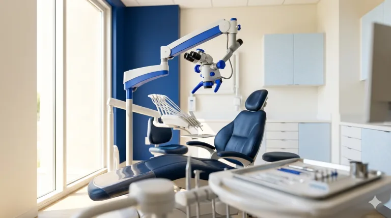
3D CBCT imaging happens during your appointment, without a separate visit and with no preparation on your part. You sit or stand while the scanner rotates once around your head; nothing touches you, and there is no tunnel to go inside. Your endodontist then reviews the images with you before treatment begins.
Find Your Nearest Office →CBCT imaging (a 3D dental scan) gives your endodontist a detailed view of the tooth, roots, and surrounding bone that a regular X-ray can't show. The scan takes seconds, uses low radiation, and helps plan precise treatment. It's especially useful for complex root canals, retreatment, and dental trauma.
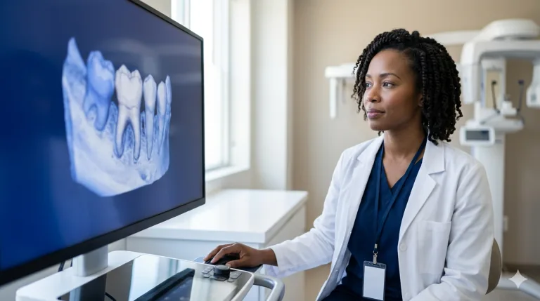
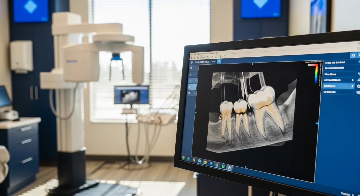
CBCT stands for cone-beam computed tomography: a quick, low-dose scan that builds a full three-dimensional model of a tooth, its roots, and the surrounding bone. A standard dental X-ray flattens all of that into a single 2D picture. The 3D view lets an endodontist map the exact canal anatomy, spot hairline cracks, and check the bone before treatment begins. At Mid-Florida Endodontics, CBCT is part of routine care for most patients because that level of detail makes treatment more precise.
Why 2D X-Rays Aren’t Enough
Standard dental X-rays have served dentistry well for over a century, but they have fundamental limitations in endodontic diagnosis. A flat image compresses a three-dimensional structure into a single plane, causing structures to overlap, hiding lesions behind dense bone. Making it impossible to determine the true number or shape of root canals from a single angle.
Conventional 2D X-Ray
- Single flat plane, structures overlap
- Misses periapical (relating to the area surrounding the very tip of a tooth’s root) lesions obscured by bone
- Cannot reveal extra canals (e.g. MB2)
- Cannot accurately assess crack depth
- Underestimates lesion size by up to 50%
- Cannot show buccolingual dimensions
3D CBCT Imaging
- Full three-dimensional visualization
- Detects periapical lesions missed on 2D
- Reveals all root canals including accessory anatomy
- Assesses crack extent and bone involvement
- Accurate lesion size and location in all dimensions
- Maps proximity to nerves, sinuses, and adjacent roots
How Mid-Florida Endodontics Uses CBCT
CBCT imaging is not a selective tool we reach for in complicated cases, it is a standard part of how we diagnose and plan every procedure. Here is what it allows us to do that would not be possible otherwise:
Identify All Root Canals Before Treatment
Upper first molars frequently have a fourth canal, the MB2, that is missed in a significant percentage of cases treated without 3D imaging. A missed canal is one of the most common causes of root canal failure. CBCT eliminates this guesswork entirely.
Detect Periapical Lesions Earlier
Research has shown CBCT detects periapical pathology significantly more often than conventional X-rays, catching bone infections at an earlier, more treatable stage when healing after treatment is faster and more predictable.
Assess Crack Depth and Extent
Cracks are largely invisible on 2D X-rays. CBCT can identify associated bone changes that indicate a crack has progressed deep enough to affect prognosis, helping us give you an accurate assessment of whether a tooth is worth treating before treatment begins.
Plan Retreatment Cases Precisely
Before retreating a previously treated tooth, CBCT reveals exactly what the original treatment missed, whether a post is present, and whether separated instruments are in the canals, allowing us to plan the procedure with full information rather than discovering complications mid-treatment.
Guide Surgical Planning
Before an apicoectomy (a minor surgical procedure that removes the tip of a tooth’s root and the surrounding infected tissue), CBCT maps the root tip position relative to the inferior alveolar nerve, maxillary sinus. Adjacent root apices, enabling surgical access that is precise, safe, and minimally invasive.
Is CBCT Safe? Understanding the Radiation Dose
Limited Field-of-View CBCT: Radiation in Context
- A limited FOV dental CBCT delivers approximately 5-50 microsieverts (μSv) depending on field size and protocol
- This is comparable to 1-6 days of natural background radiation we all receive from the environment
- A cross-country flight exposes a passenger to approximately 30-40 μSv, similar to or more than a small FOV CBCT
- A standard chest X-ray delivers approximately 100 μSv, well above a limited FOV dental CBCT
- A full-mouth series of conventional dental X-rays delivers a comparable or higher cumulative dose
The American Association of Endodontists, the American Academy of Oral and Maxillofacial Radiology, and major international dental imaging organizations all endorse CBCT use in endodontics when diagnostic benefit is present. The diagnostic value of three-dimensional imaging before treatment consistently and significantly outweighs the minimal radiation involved. (Pauwels R, et al. Effective dose range for dental cone-beam computed tomography (CBCT), a 3D X-ray that creates a complete three-dimensional image of the tooth and surrounding bone scanners. Eur J Radiol. 2012;81(2):267-271.)
What Should I Expect During a CBCT Scan?
The scan itself takes less than a minute. You will be positioned seated or standing while the CBCT unit rotates around your head in a single arc. You do not go inside a tunnel as you would with a hospital CT scanner. The machine does not touch you. You simply hold still for the duration of the rotation, most patients describe it as entirely uneventful.
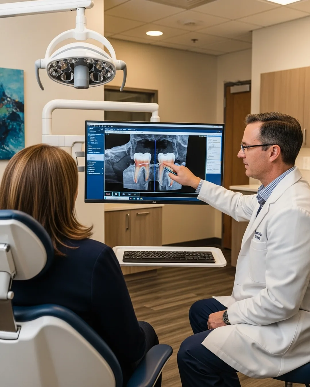
The resulting three-dimensional images are available within minutes and are reviewed by your endodontist before treatment begins at the same appointment. No special preparation is needed.
Frequently asked questions
What is a CBCT scan?
How long does a CBCT scan take?
Is CBCT imaging safe?
Why do I need a 3D scan instead of a regular X-ray?
How much does a CBCT scan cost?
Do I need a referral for CBCT imaging?
Will the CBCT scan be uncomfortable?
Can children have CBCT scans?
See what 2D X-rays miss.
3D imaging in seconds at every office. No extra visits, no referral X-rays required.
Book at your nearest officeSpecialist 3D CBCT Imaging in Endodontics near you
-
3D CBCT Imaging in Endodontics in Clermont
-
3D CBCT Imaging in Endodontics in Daytona Beach
-
3D CBCT Imaging in Endodontics in Dr. Phillips, Orlando
-
3D CBCT Imaging in Endodontics in Kissimmee
-
3D CBCT Imaging in Endodontics in Lake Nona, Orlando
-
3D CBCT Imaging in Endodontics in Longwood
-
3D CBCT Imaging in Endodontics in Maitland
-
3D CBCT Imaging in Endodontics in Mount Dora
-
3D CBCT Imaging in Endodontics in Orange City
-
3D CBCT Imaging in Endodontics in Waterford Lakes, Orlando
-
3D CBCT Imaging in Endodontics in Winter Garden