Tooth pain is one of the most diagnostically challenging symptoms in all of healthcare. It can be sharp or dull, constant or intermittent, easy to locate or seemingly everywhere. It can originate from inside a tooth, from the supporting structures, from the jaw muscles, from the sinuses, or from nerve pathways that refer pain to entirely the wrong location.
Because no single sign or test is conclusive on its own, endodontists rely on a structured sequence of tests and then weigh the findings together. Endodontic specialists are trained specifically in this diagnostic challenge. Here is exactly how they work through it, step by step.
The Endodontic Diagnostic Process
Detailed Patient History
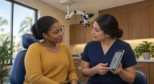
The diagnostic process begins before any instrument touches the tooth. The endodontist takes a thorough history: when did the pain start, what triggers it, how long does it last, is it getting better or worse, is it constant or intermittent, does anything relieve it, has there been any prior treatment on this tooth? This narrative guides the entire clinical examination and often provides the most important diagnostic clues before a single test is performed. Patient-reported symptoms alone do not reliably reveal the exact pulp condition, which is why the history sets up the testing that follows rather than replacing it.2
Cold Testing
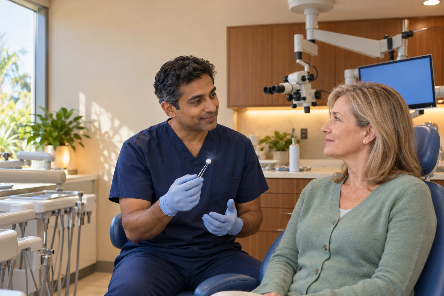
A cold stimulus, typically a refrigerant spray applied to a small cotton pellet, is applied to each tooth under evaluation and to adjacent and opposing teeth for comparison. The endodontist carefully notes whether a response is present, how intense it is, and critically, how long it lingers after the cold is removed. A response that persists after the cold is removed indicates the pulp is inflamed, and a stronger, longer-lasting response generally points to more advanced inflammation. No response at all may indicate pulp necrosis (death of the living tissue inside the tooth). Cold testing is a useful guide rather than a stand-alone verdict, so its result is always read alongside the other findings.2
Heat Testing
Heat testing is performed selectively, particularly when a patient reports that heat triggers or worsens their pain, or when cold testing results are inconclusive. A warm gutta-percha (a natural rubber-like material used to fill and seal root canals after treatment, the gold standard for over 150 years) stick or heated instrument is applied briefly to the tooth surface. Pain that lingers or intensifies with heat is a specific indicator of irreversible pulpitis (severe inflammation of the tooth’s inner tissue, too damaged to heal on its own) or advanced pulp necrosis. In some patients, heat actually relieves pain temporarily, a paradoxical finding associated with pulp necrosis and abscess formation. Pooled diagnostic-accuracy data confirm that thermal tests, cold and heat together, give endodontists meaningful information about pulp status when read as part of the full examination.6
Percussion Testing
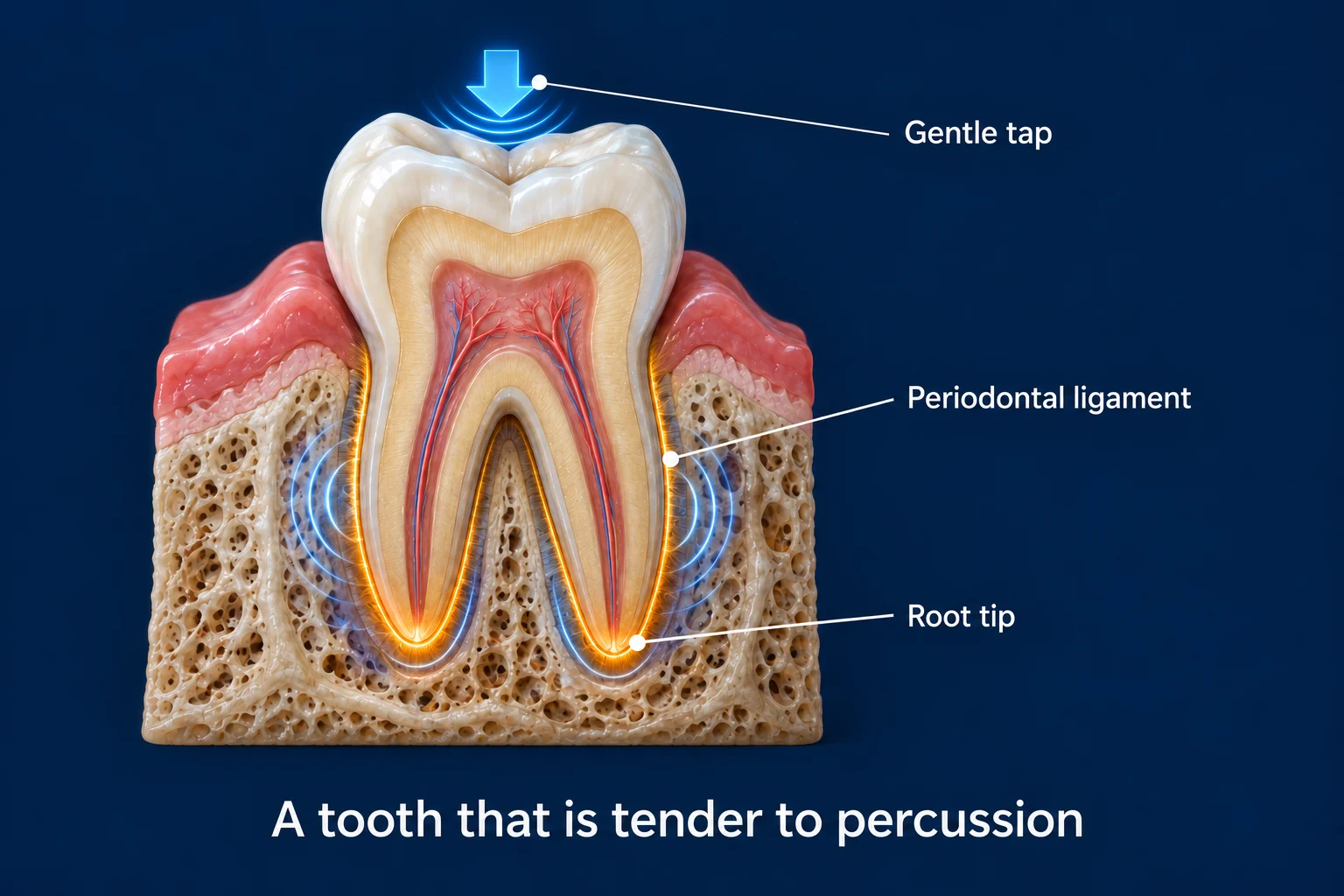
The endodontist gently taps each tooth with the handle of a dental instrument. A tooth that is tender to percussion, even light tapping, has inflammation in the periodontal ligament (the thin layer of fibers connecting the tooth root to the jawbone, which acts as a shock absorber and contains pressure-sensitive nerve fibers) surrounding the root, often indicating that infection or inflammation has spread beyond the pulp to the periapical (relating to the area surrounding the very tip of a tooth’s root) tissues. Percussion sensitivity helps confirm which tooth is the primary problem and indicates the severity of periapical involvement.
Palpation Testing
The endodontist presses on the gum tissue overlying the root tips of the teeth under evaluation. Tenderness to palpation in the apical region indicates periapical inflammation or abscess. Swelling, fluctuance (a fluid-filled sensation), or a visible sinus tract (pimple-like bump) in this area confirms active infection that has broken through the bone into soft tissue.
Bite Testing (Tooth Slooth / Bite Stick)
The patient bites down on a small plastic device one cusp at a time. Pain triggered on a specific cusp, particularly pain that occurs when releasing bite pressure rather than during it, is a classic sign of a cracked tooth. This test localizes the crack to a specific cusp and helps plan treatment. It also differentiates cracked tooth syndrome from other causes of bite pain that produce a different pattern of discomfort.
Periodontal Probing
A periodontal probe is used to measure the depth of the gum pockets around each tooth. Deep, isolated probing depths, particularly narrow, isolated pockets rather than generalized gum disease, are associated with vertical root fractures. This finding, combined with imaging, helps identify fractures that have reached the root and may affect the tooth’s prognosis. Because a probing pattern can point to more than one cause, the endodontist confirms it against imaging before reaching a conclusion.
Selective Anesthesia
When pain is poorly localized, particularly when patients cannot identify which tooth hurts, or when pain appears to refer from one jaw to the other, selective anesthesia is used. The endodontist anesthetizes specific teeth or nerve blocks in a systematic sequence. When a particular injection eliminates the pain, the source has been identified. This technique is particularly valuable for resolving diagnostic uncertainty in complex or atypical presentations.

3D CBCT Imaging
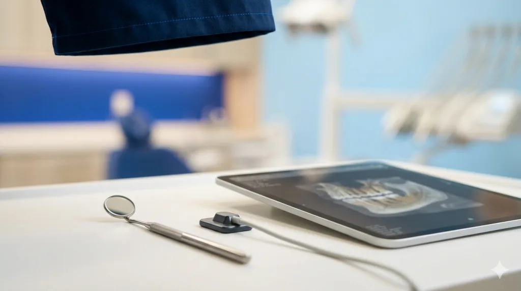
Three-dimensional cone-beam CT imaging is used routinely at Mid-Florida Endodontics on the vast majority of patients. It reveals periapical lesions that 2D X-rays can miss,4 helps identify root fractures through associated bone loss patterns, and maps canal anatomy in full three dimensions. Used by the endodontist, it provides confirmation of findings suspected from clinical testing and often changes the treatment plan in more complex cases.3 In studies where trained endodontists read both image types, they detected periapical lesions more accurately with CBCT than with conventional periapical radiographs.7 The diagnostic information it provides is fundamentally different in quality and completeness from conventional flat radiographs.
Surgical Microscope Examination
For cases where a crack is suspected but not confirmed, or where access cavity findings need to guide diagnosis, the endodontist uses the surgical microscope for magnification and illumination that reveals crack lines, pulp chamber (the hollow space inside the crown of the tooth that houses the nerve and blood vessels) anatomy, and tissue condition at a level impossible with the naked eye. In some cases, what the endodontist sees under the microscope is itself diagnostic, confirming a crack or identifying a finding that resolves a long-standing diagnostic puzzle.
How Findings Are Combined Into a Diagnosis
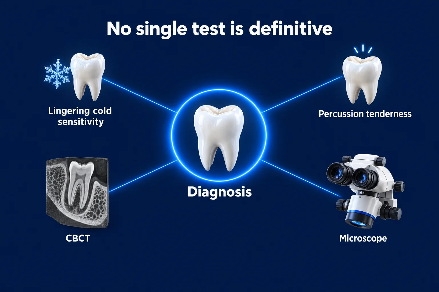
No single test is definitive in isolation. Endodontic diagnosis works by triangulating multiple findings, which is exactly why so many tests are run and why each is weighed against the others.2 A tooth with lingering cold sensitivity, percussion tenderness, and a periapical radiolucency on CBCT has a high-confidence diagnosis of symptomatic apical periodontitis (infection and inflammation at the root tip spreading into the jawbone) from irreversible pulpitis or necrosis. A tooth with bite pain on a specific cusp, rebound tenderness on releasing the bite, and a crack visible under the microscope has a high-confidence diagnosis of cracked tooth syndrome.
The diagnostic classification system used by endodontists, established by the American Association of Endodontists, defines specific pulp and periapical diagnoses based on the combination of clinical and radiographic findings.1 This structured approach ensures consistent, evidence-based treatment decisions and gives every tooth a defined diagnostic label that maps to a specific course of care. Accurate diagnosis matters for the result: the clinical condition of the tooth before treatment is among the factors most strongly linked to how well root canal treatment turns out.5
| Diagnosis | Key Findings | Treatment |
|---|---|---|
| Normal Pulp | Mild cold response, resolves quickly; no percussion tenderness; no X-ray changes | Monitor; address any underlying cause |
| Reversible Pulpitis | Brief cold sensitivity; no spontaneous pain; no periapical changes | Remove irritant; restorative treatment; monitor |
| Irreversible Pulpitis | Lingering cold/heat sensitivity; may have spontaneous pain; periapical may be normal | Root canal treatment |
| Pulp Necrosis | No cold response; possible percussion tenderness; periapical changes on imaging | Root canal treatment |
| Symptomatic Apical Periodontitis | Percussion tender; possible swelling; periapical lesion (an area of infection and bone damage at the tip of a tooth’s root, visible on X-rays) on imaging | Root canal treatment, urgent |
| Acute Apical Abscess | Severe percussion pain; swelling; possible fever; periapical lesion | Root canal treatment, same day |
The diagnostic classification used in endodontics is built on a structured set of terms defined through a systematic review of the literature by the American Association of Endodontists.1 Systematic reviews of pulp testing report that no single sign, symptom, or thermal test is accurate enough to stand alone, which is the evidence-based reason endodontists combine several tests and weigh them together rather than relying on any one result.2 For imaging, the evidence shows that 3D CBCT supplies information beyond conventional 2D radiographs and meaningfully influences endodontic diagnosis and treatment planning in more complex cases.3
Tooth pain that is hard to pinpoint is exactly what endodontists are trained for. Book an evaluation at your nearest MFE location. Find a location near you.
Works Cited
- Levin LG, Law AS, Holland GR, Abbott PV, Roda RS. Identify and define all diagnostic terms for pulpal health and disease states. J Endod. 2009;35(12):1645-1657. doi:10.1016/j.joen.2009.09.032 Systematic Review
- Mejàre IA, Axelsson S, Davidson T, et al. Diagnosis of the condition of the dental pulp: a systematic review. Int Endod J. 2012;45(7):597-613. doi:10.1111/j.1365-2591.2012.02016.x Systematic Review
- Tay KX, Lim LZ, Goh BKC, Yu VSH. Influence of cone beam computed tomography on endodontic treatment planning: a systematic review. J Dent. 2022;127:104353. doi:10.1016/j.jdent.2022.104353 Systematic Review
- Petersson A, Axelsson S, Davidson T, et al. Radiological diagnosis of periapical bone tissue lesions in endodontics: a systematic review. Int Endod J. 2012;45(9):783-801. doi:10.1111/j.1365-2591.2012.02034.x Systematic Review
- Ng YL, Mann V, Rahbaran S, Lewsey J, Gulabivala K. Outcome of primary root canal treatment: systematic review of the literature. Part 2. Influence of clinical factors. Int Endod J. 2008;41(1):6-31. doi:10.1111/j.1365-2591.2007.01323.x Systematic Review
- Patro S, Meto A, Mohanty A, et al. Diagnostic accuracy of pulp vitality tests and pulp sensibility tests for assessing pulpal health in permanent teeth: a systematic review and meta-analysis. Int J Environ Res Public Health. 2022;19(15):9599. doi:10.3390/ijerph19159599 Systematic Review
- Campello AF, Gonçalves LS, Guedes FR, Marques FV. Cone-beam computed tomography versus digital periapical radiography in the detection of artificially created periapical lesions: a pilot study of the diagnostic accuracy of endodontists using both techniques. Imaging Sci Dent. 2017;47(1):25-31. doi:10.5624/isd.2017.47.1.25 Prospective Study