
When you come to Mid-Florida Endodontics, one of the first things our specialists may do is take a CBCT scan. Patients often ask: why isn’t a regular X-ray enough? The answer reveals something fundamental about the limitations of conventional dental imaging, and why 3D imaging has become an essential tool in modern endodontic practice. More on why endodontists are specialists in finding pain other dentists miss.
How CBCT Is Different from a Standard Dental X-Ray
Standard 2D Dental X-Ray
- Flat, two-dimensional projection
- Structures overlap and obscure each other
- Cannot show depth or exact location
- Can miss periapical lesions that are visible in three dimensions
- Cannot reliably show cracks
- Cannot count or map all canals in complex teeth
- Low radiation dose
- Fast and inexpensive
3D CBCT Imaging
- True three-dimensional volumetric data
- Structures viewed in any plane without overlap
- Precise depth, width, and location measurement
- Helps the endodontist detect periapical lesions earlier
- Can reveal crack-related bone changes in some cases
- Maps full canal anatomy including extra canals
- Higher radiation than 2D (small field of view used)
- Takes seconds; reconstruction in minutes
What Can CBCT Reveal That Regular X-Rays Cannot?
Periapical Infections (Abscesses)
Research comparing the two methods has shown that standard 2D radiographs can miss periapical lesions, infections at the root tip, that are clearly visible to the endodontist on CBCT.1 When we see a lesion earlier, that can mean earlier treatment, less bone destruction, and better healing outcomes. CBCT is also valuable for confirming lesion healing at follow-up appointments, where it has been used to assess endodontic treatment results in detail.4
Canal Anatomy and Extra Canals
Some teeth have more canals than textbooks describe. Upper first molars frequently have a fourth canal (MB2) that is easily missed on 2D X-rays but far more visible to the endodontist on CBCT. Lower incisors often have two canals. C-shaped canal systems in lower molars are notoriously complex. Identifying all canals before treatment begins, rather than discovering missed canals at retreatment, is one of CBCT’s greatest clinical contributions. Because a flat projection overlaps and distorts these structures, three-dimensional imaging gives us anatomy that a single 2D film cannot reliably show.2
Root and Tooth Fractures
Vertical root fractures, cracks running along the length of the root, are among the most difficult conditions to diagnose in dentistry. They are often invisible on standard X-rays until bone loss has progressed. On CBCT, we can sometimes see characteristic halo-shaped bone loss patterns adjacent to root fractures, which helps guide diagnosis and treatment planning. We break down how endodontists use imaging to diagnose tooth pain.
Root Canal Curvature and Length
Root canals are three-dimensional structures. A canal that appears relatively straight on a 2D X-ray may have a significant buccolingual (front-to-back) curve invisible from the side view.2 CBCT reveals the true three-dimensional path of each canal, allowing the endodontist to select instruments and techniques matched to the actual anatomy rather than what a flat projection suggests.
Bone Loss Assessment
Periapical bone destruction is three-dimensional, it extends in all directions from the root tip. Standard X-rays show only the mesiodistal (side-to-side) extent of bone loss. CBCT reveals the full three-dimensional extent, which affects treatment planning, including whether non-surgical root canal treatment is sufficient or whether surgical intervention may be needed. This kind of added information has been shown to change endodontic treatment decisions in a meaningful share of cases.3
Resorption and Root Anomalies
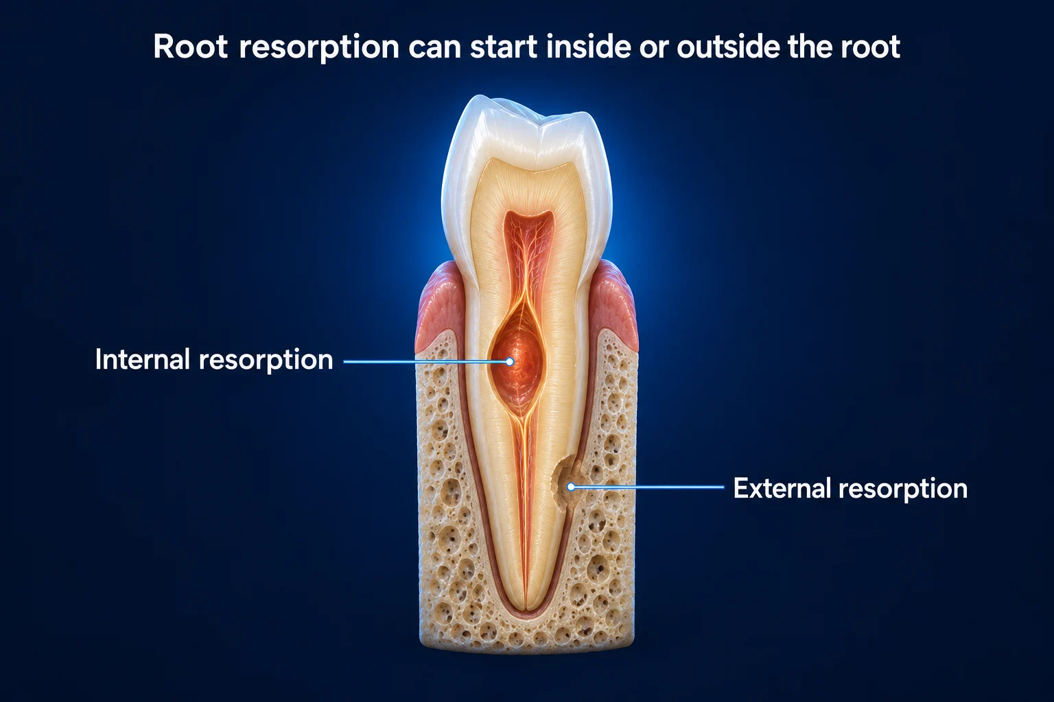
Internal or external root resorption (a process where the body’s own cells begin breaking down tooth structure, which can affect the root from the inside or outside, and can occur after trauma or due to other causes) is far more accurately assessed on CBCT than on 2D X-rays. Understanding the extent and location of resorption is critical to determining whether a tooth is treatable or whether extraction may be the better option for long-term oral health.
Is CBCT Safe? Understanding the Radiation Dose
CBCT Radiation: Kept in Perspective
A limited field-of-view dental CBCT, the type used in endodontics, delivers a radiation dose that is low in the context of everyday life. A phantom dosimetry study measuring a wide range of CBCT scanners reported effective doses spanning roughly 19 to 368 microsieverts (μSv) across all field sizes and protocols, with the smallest, most focused fields at the low end of that range.5 The dose is strongly tied to field size, so a small field of view targeted to a single tooth keeps exposure toward the lower end. For context:
- A limited field-of-view dental CBCT, focused on a single tooth or small region, uses one of the lowest-dose CBCT protocols available, because dose scales with the size of the scanned field.5
- Effective dose varies widely by device, exposure settings, and field size, which is why protocol selection matters as much as the machine itself.5
- The dose should always be weighed against the diagnostic value of the image, since image-quality needs differ from patient to patient.5
- Choosing the smallest field of view that answers the clinical question is a core principle of dose optimization in dental CBCT.5
- Because the scan is selected only when it will guide care, the small added exposure is balanced by clearer, more confident treatment planning.
The American Association of Endodontists and the American Academy of Oral and Maxillofacial Radiology endorse CBCT use in endodontics when the diagnostic benefit is present, which, in a specialty dedicated to precise diagnosis of internal tooth structure, it routinely is.6 The diagnostic value of seeing a tooth’s internal anatomy in three dimensions before treatment is weighed against the radiation involved, and we use the smallest field of view that answers the clinical question. See how endodontists use dental microscopes alongside imaging.
When Will My Endodontist Use CBCT?
At Mid-Florida Endodontics, CBCT imaging is used for the majority of patients when the diagnostic benefit is present. The three-dimensional information it provides is often too valuable to forgo: it reveals canal anatomy, periapical lesions, and bone changes, along with fractures that 2D X-rays can miss, and that missing information can change the entire diagnosis and treatment plan.3 Seeing the tooth in three dimensions before treatment begins is a fundamental part of how we deliver accurate, confident endodontic care.
Specific situations where CBCT is particularly critical include:
- Initial diagnosis, confirming the source of tooth pain and identifying periapical pathology
- Complex multi-rooted teeth where canal number and anatomy need full mapping
- Suspected or confirmed root fracture
- Root resorption assessment
- Retreatment planning, understanding existing materials and missed canals
- Traumatic dental injuries requiring bone and root assessment
- Pre-surgical endodontic planning, identifying proximity to nerves, sinus floors, and adjacent roots
- Any case where 2D imaging leaves clinical questions unanswered
The value of CBCT over conventional radiography for endodontic applications is well documented. A retrospective comparison of limited cone-beam CT against intraoral periapical radiography found that the 3D technique revealed periapical lesions in additional teeth and roots that the periapical radiographs did not show, and that observers agreed it added clinically relevant information in most cases.1 A narrative review of endodontic imaging similarly described the inherent limitations of two-dimensional periapical films, overlap, geometric distortion, and anatomical noise, and explained how CBCT helps overcome them.2 A systematic review of how CBCT influences endodontic treatment planning reported that additional CBCT information changed clinicians’ treatment plans in a substantial share of cases, particularly in higher-difficulty situations, evaluation of periapical healing, and pre-surgical planning.3 Regarding radiation safety, a phantom dosimetry study measuring effective doses across many CBCT scanners found that dose is strongly related to field size, supporting the use of the smallest field of view that answers the clinical question and reserving the scan for cases where the diagnostic benefit is present.5 Professional guidance from the American Association of Endodontists and the American Academy of Oral and Maxillofacial Radiology aligns with this benefit-driven approach to selecting CBCT in endodontics.6
MFE uses CBCT imaging at most locations across Florida as a standard part of the diagnostic process. Find your nearest location.
Works Cited
- Lofthag-Hansen S, Huumonen S, Gröndahl K, Gröndahl HG. Limited cone-beam CT and intraoral radiography for the diagnosis of periapical pathology. Oral Surg Oral Med Oral Pathol Oral Radiol Endod. 2007;103(1):114-119. doi:10.1016/j.tripleo.2006.01.001 Prospective Study
- Patel S, Dawood A, Whaites E, Pitt Ford T. New dimensions in endodontic imaging: part 1. Conventional and alternative radiographic systems. Int Endod J. 2009;42(6):447-462. doi:10.1111/j.1365-2591.2008.01530.x
- Tay KX, Lim LZ, Goh BKC, Yu VSH. Influence of cone beam computed tomography on endodontic treatment planning: a systematic review. J Dent. 2022;127:104353. doi:10.1016/j.jdent.2022.104353 Systematic Review
- Brochado Martins JF, Georgiou AC, Diogo Nunes P, et al. CBCT-assessed outcomes and prognostic factors of primary endodontic treatment and retreatment: a systematic review and meta-analysis. J Endod. 2025;51(6):687-706. doi:10.1016/j.joen.2025.03.004 Systematic Review
- Pauwels R, Beinsberger J, Collaert B, et al. Effective dose range for dental cone beam computed tomography scanners. Eur J Radiol. 2012;81(2):267-271. doi:10.1016/j.ejrad.2010.11.028
- American Association of Endodontists; American Academy of Oral and Maxillofacial Radiology. AAE and AAOMR joint position statement: use of cone beam computed tomography in endodontics 2015 update. J Endod. 2015;41(9):1393-1396. doi:10.1016/j.joen.2015.07.013