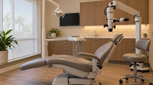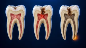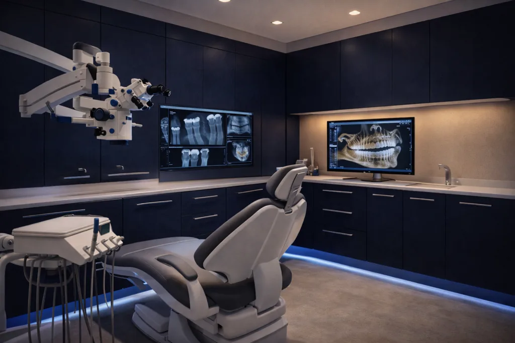
The human eye, even under excellent lighting, can resolve detail down to about 0.2 millimeters. A root canal orifice may be a fraction of a millimeter wide. A hairline crack in dentin (the layer of tooth beneath the hard enamel (the hard outer shell of the tooth, the hardest substance in the human body), softer and more sensitive, containing microscopic channels that connect to the nerve) is thinner than a human hair. Without magnification, an endodontist is working at or beyond the limits of unaided human vision on every single case.7 The microscope brings that work back inside the range the human eye can manage, so the endodontist can see clearly what is otherwise only felt or inferred.
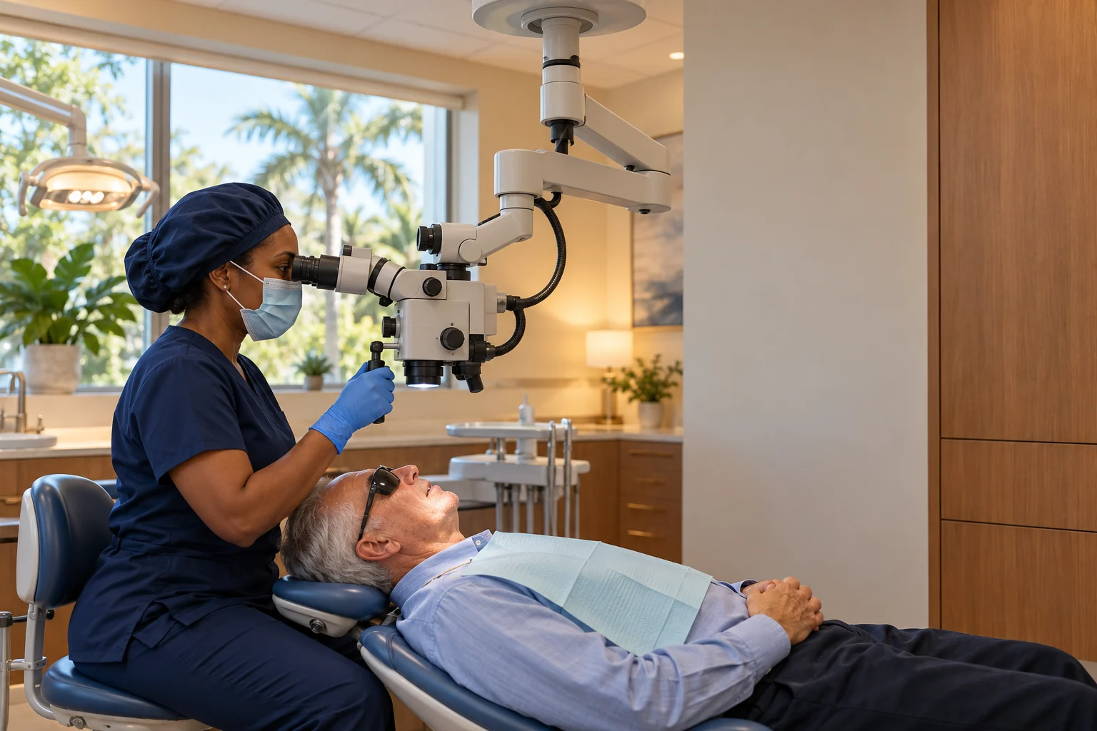
The surgical operating microscope changed endodontics fundamentally, not as a luxury addition to well-equipped practices, but as a core diagnostic and treatment tool that raises the standard of care for every patient who sits in the chair. It supports the clinician at each step, from locating canals to confirming a canal is clean before it is sealed. That care is part of why a root canal can save a tooth.
What the Microscope Enables That the Naked Eye Cannot
Identifying All Root Canals
Many teeth have more canals than standard anatomy charts suggest. Upper first molars frequently have a fourth canal, the MB2, and it is missed far more often when the endodontist works without magnification. In a systematic review and meta-analysis, the second mesiobuccal canal was located by direct vision alone in about 27% of upper first molars, rising to roughly 60% when the endodontist used magnification, and to about 72% when magnification was combined with ultrasonic instrumentation.1 Under the microscope, we read the floor of the pulp (the soft living tissue inside the tooth, containing nerves and blood vessels) chamber (the hollow space inside the crown of the tooth that houses the nerve and blood vessels), the canal orifice patterns, and subtle color variations in the dentin that guide us to every canal that needs treatment. A missed canal left untreated is one of the most common causes of root canal failure and the need for retreatment.
Detecting Cracks and Fractures
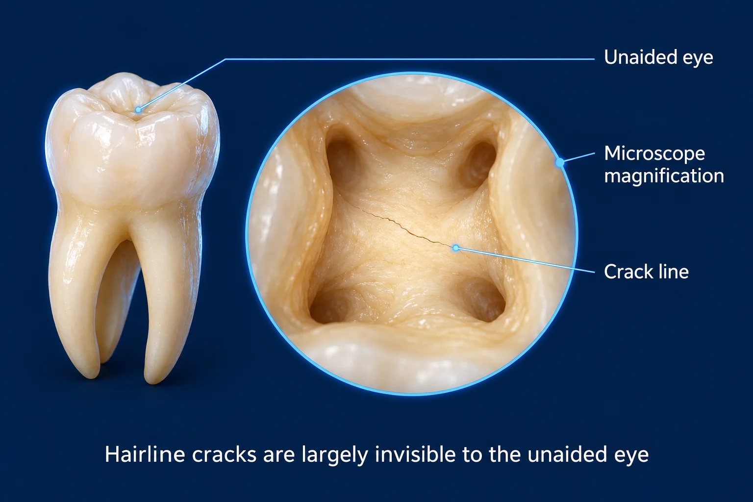
Hairline cracks in tooth structure are largely invisible to the unaided eye, and on X-rays. Under microscope magnification with coaxial lighting, crack lines often become visible as fine interruptions in the reflective surface of the dentin, or as stained lines in the pulp floor. This allows us to identify cracks that explain a patient’s symptoms even when all other findings are inconclusive, and to assess whether a crack extends into the root where it would compromise the tooth’s prognosis.
Complete Pulp Tissue Removal
Residual pulp tissue left in a canal, particularly in accessory canals or canal fins, can become a source of persistent inflammation or reinfection. Under the microscope, we can directly visualize the canal walls to confirm that tissue has been fully removed and that the canal is clean before obturation (the step where cleaned root canals are filled and sealed to prevent bacteria from re-entering). This direct visual confirmation is not possible without magnification, and it lets us treat each canal to the same clear standard.
Removing Separated Instruments
Occasionally, a file tip can separate and become lodged inside a narrow canal. Under microscope magnification, we can locate these fragments and often retrieve them using ultrasonic instruments, a procedure that is essentially impossible without both the visualization the microscope provides and the precision of ultrasonic tips. In a prospective clinical study, the success rate for removing or bypassing a fractured instrument was about 85% when the fragment was visible under the operating microscope, compared with about 48% when it was not, roughly double the success when the endodontist could see the fragment.2 In cases where removal is not feasible, the microscope helps us assess whether the fragment can be bypassed or whether it affects the overall prognosis. Here’s what happens step by step during a root canal procedure.
Endodontic Microsurgery (Apicoectomy)
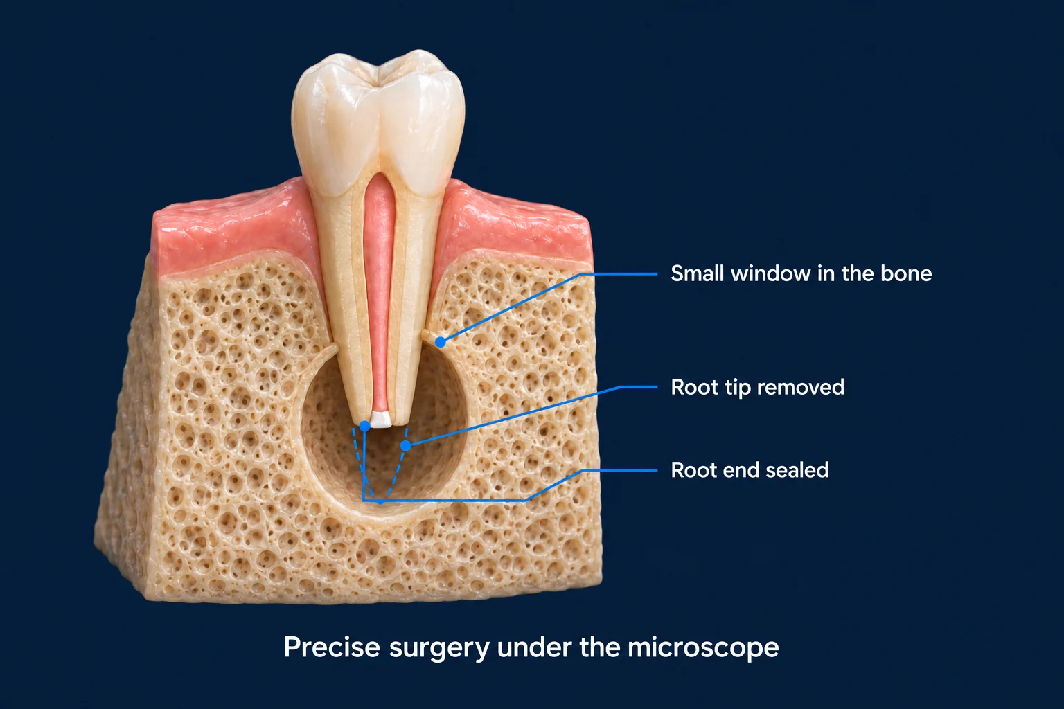
When surgical access to the root tip is required, as in an apicoectomy (a minor surgical procedure that removes the tip of a tooth’s root and the surrounding infected tissue), the microscope is indispensable. The surgical field is deep, narrow, and millimeters from vital structures. Microscope-assisted endodontic surgery allows precise root-end resection (surgically removing the very tip of the root during an apicoectomy) and retrofill (sealing the cut root end from the outside during apicoectomy surgery) preparation that is difficult to achieve at the level of accuracy needed without magnification. In a meta-analysis of surgical outcomes, the pooled success rate for endodontic microsurgery was about 94%, compared with about 59% for the older traditional root-end surgery approach.3 A prospective clinical study of surgeries performed under the operating microscope reported a comparable success rate of about 93%.4
Ultrasonic Instrumentation
Ultrasonics are a versatile and essential tool in endodontics, used for far more than irrigation. Under microscope guidance, we use ultrasonic tips to perform troughing (using an ultrasonic instrument to carefully remove a thin groove of tooth structure to locate hidden root canal openings) and access refinement to locate hidden canal orifices, to retrieve separated instruments lodged in canals, and to remove posts and other restorative materials during retreatment. We also activate irrigating solutions to support thorough disinfection throughout the canal system. The combination of microscope visualization and ultrasonic precision makes these procedures reliably achievable in skilled hands.
Microscope vs. No Microscope: What the Difference Looks Like
Without Microscope
- Canal orifices located by feel and standard anatomy knowledge
- Cracks often undetectable
- Missed canals more likely in complex anatomy
- Residual tissue removal less verifiable
- Separated instrument removal extremely difficult
- Surgery requires larger access and less precision
With Surgical Microscope
- Direct visualization of all canal orifices
- Crack lines often visible under coaxial illumination
- Extra canals reliably identified
- Clean canal walls confirmed visually before sealing
- Separated instruments frequently retrievable
- Microsurgery with minimal tissue disruption
Is the Microscope Used on Every Patient at Mid-Florida Endodontics?
Yes. The surgical operating microscope is used on every root canal, retreatment, and surgical case at Mid-Florida Endodontics, not selectively, but as a standard of practice. Just as a 3D CBCT scan provides diagnostic information that a flat X-ray cannot,5 the microscope provides visual information that unaided vision cannot. Using these tools on every patient reflects a commitment to the highest achievable standard of care, not an add-on for complex cases only. Understand what CBCT imaging adds to endodontic diagnosis.
The impact of magnification on endodontic care is documented in the literature. A systematic review and meta-analysis found that locating the second mesiobuccal (MB2) canal in upper first molars was far more reliable with magnification than with direct vision alone, and more reliable still when magnification was paired with ultrasonic instrumentation.1 A separate meta-analysis of surgical outcomes reported that endodontic microsurgery, performed under the operating microscope, achieved a markedly higher pooled success rate than traditional root-end surgery,3 a finding echoed by prospective clinical studies of microscope-assisted surgery.4 In each case the evidence supports the same point: with better visualization, the endodontist sees more, treats more completely, and works with greater precision. Outcome studies using cone-beam CT continue to refine how we measure these results.6
For more information, see why endodontists are specialists in finding problems other dentists miss.
Microscope-assisted endodontics is standard at MFE locations across Florida. Find your nearest location.
Works Cited
- Anirudhan S, Suneelkumar C, Uppalapati H, Anumula L, Kirubakaran R. Detection of second mesiobuccal canals in maxillary first molars of the Indian population: a systematic review and meta-analysis. Evid Based Dent. 2022. doi:10.1038/s41432-022-0233-3 Systematic Review
- Nevares G, Cunha RS, Zuolo ML, Bueno CE. Success rates for removing or bypassing fractured instruments: a prospective clinical study. J Endod. 2012;38(4):442-444. doi:10.1016/j.joen.2011.12.009 Prospective Study
- Setzer FC, Shah SB, Kohli MR, Karabucak B, Kim S. Outcome of endodontic surgery: a meta-analysis of the literature. Part 1: comparison of traditional root-end surgery and endodontic microsurgery. J Endod. 2010;36(11):1757-1765. doi:10.1016/j.joen.2010.08.007 Systematic Review
- Song M, Shin SJ, Kim E. Outcomes of endodontic micro-resurgery: a prospective clinical study. J Endod. 2011;37(3):316-320. doi:10.1016/j.joen.2010.11.029 Prospective Study
- Tay KX, Lim LZ, Goh BKC, Yu VSH. Influence of cone beam computed tomography on endodontic treatment planning: a systematic review. J Dent. 2022;127:104353. doi:10.1016/j.jdent.2022.104353 Systematic Review
- Brochado Martins JF, Georgiou AC, Nunes PD, et al. CBCT-assessed outcomes and prognostic factors of primary endodontic treatment and retreatment: a systematic review and meta-analysis. J Endod. 2025;51(6):687-706. doi:10.1016/j.joen.2025.03.004 Systematic Review
- Chauhan S, Chauhan R, Bhasin P, Bhasin M. Magnification: the game changer in dentistry. World J Methodol. 2025;15(2):100937. doi:10.5662/wjm.v15.i2.100937 Narrative Review