
One of the most important and often misunderstood aspects of dental trauma is that it is not a single event, it is the beginning of a monitoring process. An injured tooth that looks fine on the day of injury may quietly develop pulp necrosis over the following weeks or months with no symptoms until an abscess forms. A root canal due to trauma is needed once the pulp becomes necrotic (dies).

Understanding when a traumatized tooth needs root canal treatment, and when it can be safely monitored, is essential for anyone who has experienced or is caring for someone with a dental injury. The international consensus guidelines that endodontic specialists follow set out, injury by injury, which teeth warrant treatment right away and which can be watched over time.1
Which Injuries Most Commonly Lead to Root Canal Treatment?
| Injury Type | Immediate Root Canal Needed? | Likely to Need Root Canal Later? | Notes |
|---|---|---|---|
| Avulsion (knocked out) | Almost always | Yes, after replantation | Root canal treatment typically initiated 7-14 days after replantation in mature teeth |
| Intrusion (pushed in) | Usually yes | High likelihood | Severe vascular disruption; pulp necrosis very common |
| Lateral luxation (displaced) | Sometimes | Moderate likelihood | Depends on severity and age of patient; monitor closely |
| Extrusion (partially out) | Sometimes | Moderate likelihood | Pulp may recover in young patients; monitor |
| Crown fracture with pulp exposure | Usually yes | Yes if pulp cap fails | Pulp capping attempted in select cases; Root canal treatment often required |
| Crown fracture, no pulp exposure | Usually not immediately | Possible over time | Monitor pulp response; may develop necrosis months later |
| Concussion (no displacement) | No | Low but possible | Monitor at 4, 8, and 12 weeks; some develop delayed necrosis |
| Root fracture (horizontal) | Sometimes | Depends on fracture location | Coronal third fractures: higher root canal need. Apical third: often heal |
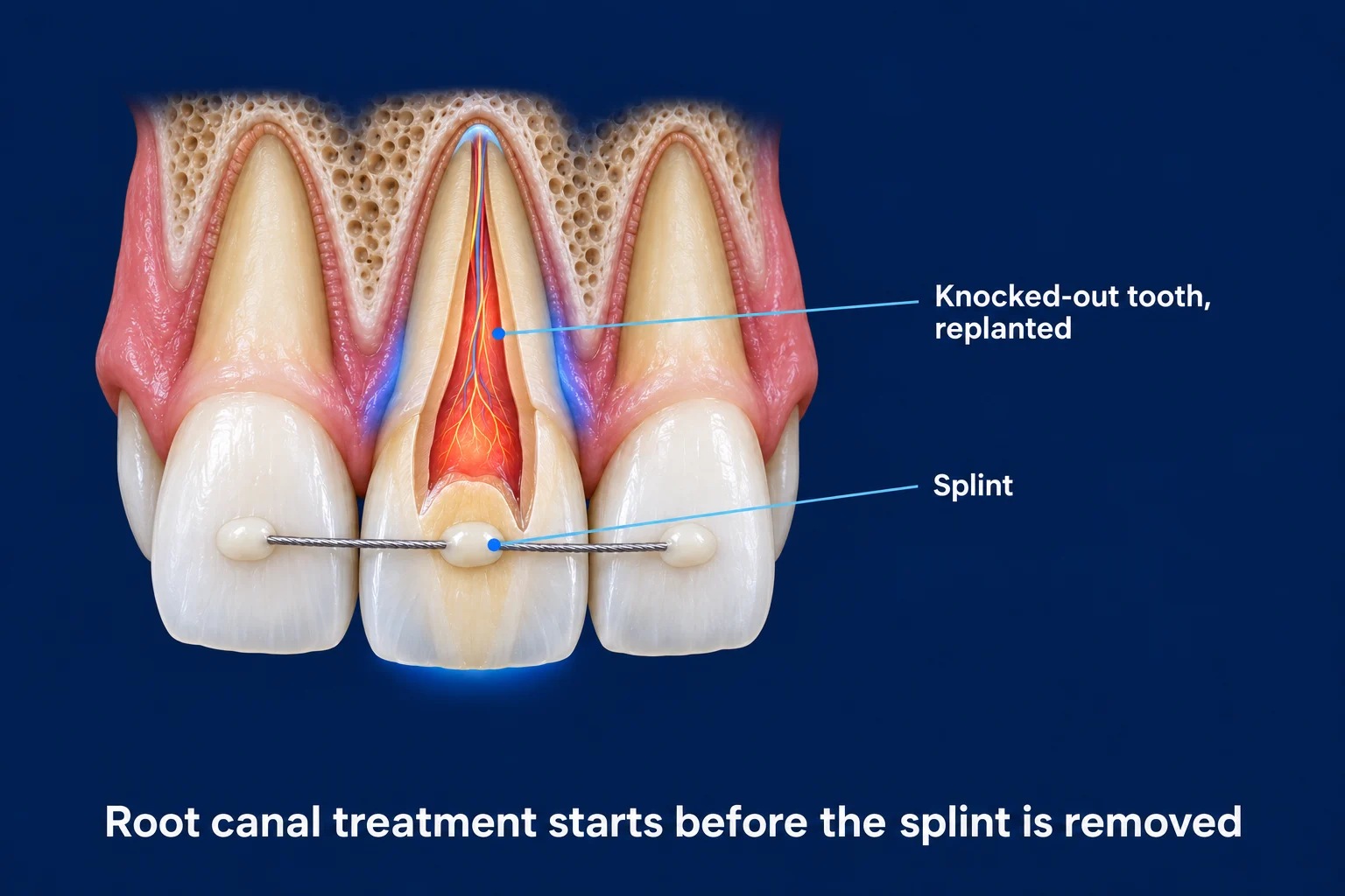
For an avulsed (knocked-out) permanent tooth that has been replanted, the international trauma guidelines recommend starting root canal treatment within about one to two weeks in a mature tooth, before the splint is removed, to prevent infection-driven root resorption.2 This is why a knocked-out adult tooth almost always needs endodontic care even when it looks healthy after being put back in place.
Signs That a Traumatized Tooth Needs Root Canal Treatment
Act Now. Contact Your Endodontist
- Pulp visibly exposed after fracture
- Spontaneous throbbing or severe pain
- Swelling developing near the tooth
- Tooth darkening to gray or brown
- Pimple-like bump on gum near the tooth
- Fever alongside dental pain
Monitor at Your Next Follow-Up
- Tooth no longer responds to cold testing
- Dark shadow appearing at root tip on X-ray
- Sensitivity that was improving has returned
- Gradual, progressive tooth darkening
- Mild persistent ache months after injury
Why Traumatized Teeth Can Fail Silently
After an injury that disrupts the blood supply to the pulp, particularly from intrusion or avulsion (when a tooth is completely knocked out of its socket, a dental emergency), the pulp tissue can die gradually without producing obvious pain. The nerve tissue degenerates, but the infection that follows may progress slowly through the root canal system and into the surrounding bone without triggering the acute throbbing pain people associate with dental infection.
This silent necrosis is why follow-up monitoring is non-negotiable after significant dental trauma. The tooth may feel perfectly fine while a chronic infection develops at the root tip. Regular pulp vitality (whether the living tissue inside the tooth is still healthy and functioning) testing and radiographic monitoring at scheduled intervals can detect this before symptoms, or bone destruction, become significant.1 When deeper imaging is needed, 3D CBCT gives the most accurate view of healing at the root tip, which is why endodontic specialists use it to judge whether a treated tooth is recovering.4
A tooth that gradually darkens after trauma, turning gray compared to adjacent teeth, is a reliable visible sign of pulp necrosis even in the absence of pain. This discoloration comes from blood breakdown products released by the dying pulp tissue. This tooth needs endodontic evaluation regardless of whether it hurts. Long-term clinical studies of luxated, non-vital incisors confirm that such teeth can be treated successfully once necrosis is recognized and managed.3
Follow-Up Schedule After Dental Trauma
Standard Monitoring Timeline After Significant Trauma
Initial evaluation, diagnosis, and any immediate treatment (splinting, pulp capping, or root canal if indicated). 3D CBCT (cone-beam CT, a low-dose 3D X-ray that lets us see the tooth and bone from every angle) imaging to assess root and bone damage.
Splint removal (if applied) and initial pulp vitality assessment. Root canal initiated for replanted avulsed teeth at this stage in most protocols.2
Pulp testing and clinical examination. Signs of pulp necrosis or periapical (relating to the area surrounding the very tip of a tooth’s root) changes begin to appear at this interval in teeth that will not recover.
Radiographic follow-up. Periapical bone changes visible if necrosis has occurred. Root canal treatment initiated if necrosis is confirmed.
Comprehensive review. Most teeth that will develop necrosis will have shown signs by this point. Confirm healing of any treated lesions.
Annual review for 2-5 years post-injury. Some teeth develop very late pulp changes. Long-term monitoring is part of complete trauma management.1
The International Association of Dental Traumatology (IADT) provides evidence-based guidelines for the management of all categories of traumatic dental injuries, including specific protocols for when root canal treatment is indicated immediately versus when monitoring is appropriate. Research supporting these guidelines consistently identifies pulp necrosis, confirmed by lack of response to vitality testing, radiographic periapical changes, and tooth discoloration, as the primary criteria triggering endodontic intervention. For avulsed permanent teeth that are replanted, the guidelines recommend initiating root canal treatment within the first one to two weeks to prevent infection-related root resorption.12 (DiAngelis AJ, Andreasen JO, Ebeleseder KA, et al. International Association of Dental Traumatology guidelines for the management of traumatic dental injuries: 1. Fractures and luxations of permanent teeth. Dent Traumatol. 2012;28(1):2-12.)
Works Cited
- DiAngelis AJ, Andreasen JO, Ebeleseder KA, et al. International Association of Dental Traumatology guidelines for the management of traumatic dental injuries: 1. Fractures and luxations of permanent teeth. Dent Traumatol. 2012;28(1):2-12. doi:10.1111/j.1600-9657.2011.01103.x
- Andersson L, Andreasen JO, Day P, et al. International Association of Dental Traumatology guidelines for the management of traumatic dental injuries: 2. Avulsion of permanent teeth. Dent Traumatol. 2012;28(2):88-96. doi:10.1111/j.1600-9657.2012.01125.x
- Cvek M. Prognosis of luxated non-vital maxillary incisors treated with calcium hydroxide and filled with gutta-percha (a natural rubber-like material used to fill and seal root canals after treatment, the gold standard for over 150 years). A retrospective clinical study. Endod Dent Traumatol. 1992;8(2):45-55. doi:10.1111/j.1600-9657.1992.tb00228.x
- Brochado Martins JF, et al. CBCT-assessed outcomes and prognostic factors of primary endodontic treatment and retreatment: a systematic review and meta-analysis. J Endod. 2025;51(6):687-706. doi:10.1016/j.joen.2025.03.004 Systematic Review